COVID-19, Flu and RSV – What makes them different?
Written by Victoria Simms, September 28, 2022. Reviewed April 24, 2024.

Despite causing similar symptoms, there are key differences between COVID-19, flu and RSV such as the size and structure of the viral genomes as well as the virion proteins. These differences allow us to design tests to distinguish between these viral infections and develop treatments and vaccines specific to each.
Respiratory infections are commonly caused by viruses and at present the three most common respiratory viruses within the UK are coronavirus (COVID-19), influenza virus (flu) and respiratory syncytical virus (RSV). These three viruses share similarities including causing similar symptoms, being highly contagious, and increased transmission in colder months, however their viral genomes and particle structures are uniquely different. These differences allow us to design tests to distinguish between viral infections that appear the same from symptoms and develop treatments and vaccines specific to each. Understanding these viruses, their genomes and mechanisms of infection helps us to reduce the rate of viral transmission and improve patient care.
Read on to learn more about each virus and how they differ.
How do COVID-19, Flu and RSV differ?
COVID-19 is caused by a coronavirus known as SARS-CoV-2 (Severe Acute Respiratory Syndrome), the SARS-CoV-2 genome of 29.9 kb is significantly larger than that of influenza and RSV at 13.5 kb and 15.2 kb respectively (see Table 1). In fact, the SARS-CoV-2 genome is currently the largest identified animal RNA virus, mainly due to its 'proof-reading' capabilities which identifies and corrects errors that arise during viral replication.1
All three viruses have single stranded RNA as their genetic material; however, the genome of influenza and RSV is negatively sensed, whereas the SARS-CoV-2 genome is positively sensed, which gives this virus the ability to function as messenger RNA and the genome can be directly translated into protein inside the host cell. Furthermore, the RNA genome of SARS-CoV-2 and RSV is unsegmented; however, the influenza genome is separated into eight single stands of RNA. This segmented genome enables antigenic shift, in which segment reassortment can occur with other circulating human and animal viruses, this can create a new strain of the virus which has novel antigenic surface proteins and gives it the capability to easily infect individuals.2
To propagate infection, COVID-19, flu and RSV produce spherical shaped virions which are transmitted within respiratory droplets, spread via direct contact, coughing or sneezing.3,4 All three viruses use epithelial cells within the respiratory tract as host cells to undergo viral replication. Once inside the host cell, the viral components are replicated, transcribed, and translated hundreds of times producing many new viral particles to propagate infection further. Gaining access to the host cells is a crucial step within the viral replication process and is highly dependent on the surface proteins, and these are specific to each virus.
Read on to find out more about how each virus is different and how we can test for them.
Table 1: Comparisons between the viral genomes of SARS-CoV-2, Influenza and RSV.
| SARS-CoV-2 (COVID-19) | 29.9 | Single stranded RNA (ssRNA) | Positive | Non-segmented | 1 | Respiratory droplets |
| Influenza (flu) | 13.5 | Single stranded RNA (ssRNA) | Negative | 8 Segments (strains A and B), 7segments (strain C) | 4 | Respiratory droplets |
| Respiratory syncytial virus (RSV) | 15.2 | Single stranded RNA (ssRNA) | Negative | Non-segmented | 2 | Respiratory droplets |
What is COVID-19?
COVID-19 is caused by a coronavirus known as SARS-CoV-2, a member of the Sarbecovirus subgenus (β-CoV lineage B) and the seventh member of the coronavirus family known to infect humans5, SARS-CoV-2 is related to the SARS-CoV-1 virus which was responsible for the 2002-2004 SARS outbreak.
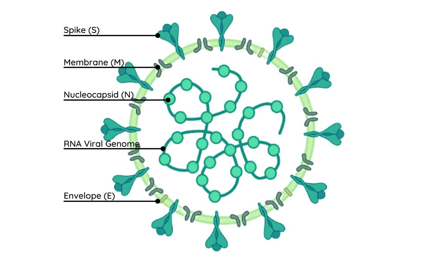
Figure 1: Virion structure of SARS-CoV-2 (COVID-19).
The viral particles of SARS-CoV-2 are 80-120 nm in size and consist of a positively sensed single-stranded RNA genome attached to nucleocapsid (N) proteins (Figure 1). The N proteins play a crucial role in viral replication and are highly conserved, which has made them a target for drug development to decrease the rate of COVID-19 transmission.1
The SARS-CoV-2 genome is surrounded by a protective lipid envelope known as a capsid, which consists of three structural proteins, membrane (M), envelope (E) and spike (S) proteins.6,7 The trimeric S glycoproteins appear as club-shaped projections on the surface of the viral particle, which gives rise to the 'corona' name of this virus, meaning 'crownlike' or 'sun'. The S proteins enable the virus to gain access into the host cells by the receptor binding domain (RBD) within S1 subunit of the protein interacting and binding to the Angiotensin Converting Enzyme 2 (ACE2) receptor on the surface of the epithelial cells.8
Inside the host cells, viral replication occurs which can introduce mutations into the 29,903 bases of the SARS-CoV-2 genome and recombination events cause genetic changes which brings about new variants. These new variants can have enhanced viral transmissibility and increase immune resistance. Since the beginning of the COVID-19 pandemic, the World Health Organization (WHO) has declared five variants a concern for human health: alpha, beta, gamma, delta and omicron.9
Genetic sequencing of COVID-19 variants has revealed high levels of mutagenesis in the S protein, this has subsequently led to amino acid alterations within the 1273 amino acid sequence and impacted S protein functionality and viral transmission; for example, the D614G mutation increases infectivity by assembling more functional glycoproteins on the virion surface, whereas the S477N mutation within the RBD increases the S protein binding affinity to ACE2 and confers resistance to antibodies, thereby accelerating the spread of SARS-CoV-2.10
The genetic sequence of the S protein has also gained interest from a diagnostic perspective as the coding sequence has been targeted in the design and development of the COVID-19 vaccinations. The Oxford-AstraZeneca COVID-19 vaccine contains the full-length coding sequence of the SARS-CoV-2 S protein within an adenovirus vector and the Pfizer and Moderna vaccines use the S protein mRNA to enable individuals to produce antibodies to recognise the SARS-CoV-2 virus if infected. The high levels of mutagenesis within the S protein poses challenges in protecting against new variants, therefore the N protein, which is much more highly conserved between different variants, has recently been utilised to protect against new variants and provide a better immune response against other coronavirus family members.
What is Flu?
Flu is caused by the influenza virus belonging to the Orthomyxoviridae family. There are three strains, A, B and C which infect humans; however, it is only Influenza A and B that cause concerns for human health.11
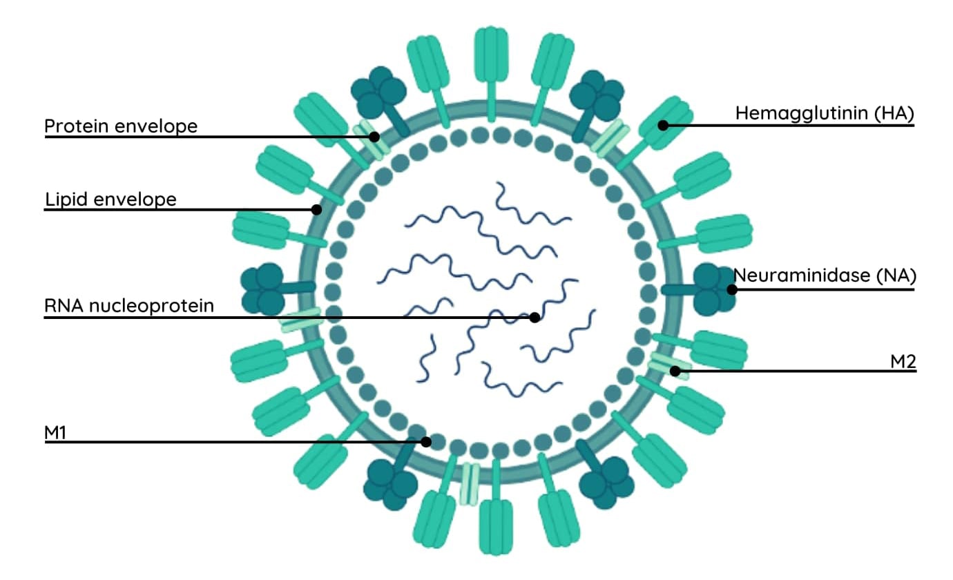
Figure 2: Virion structure of Influenza.
Influenza virions are typically spherical or ovoid shaped with a diameter of around 100 nm, but they can also form filamentous structures with lengths of around 300 nm. The genetic material of the influenza virus is RNA of size 13.5 kb, it contains eight genes and encodes for 11 different proteins. Unlike the coronavirus genome, the influenza genome is negatively sensed and segmented; strains A and B are comprised of eight single stranded RNA segments, whereas strain C has 7 RNA segments.12 The RNA genome exists in the centre of the virion (Figure 2) and is attached to nucleoproteins to form a helical ribonucleoprotein, which is encapsulated by a lipid bilayer.13
Influenza virions have several unique surface proteins which function to enable the virus to bind and replicate efficiently inside the host cell. Hemagglutinin (HA) is the most abundant glycoprotein on the surface of the influenza virions, HA makes up approximately 80% of the surface proteins and is responsible for interacting and binding to sialic acid on the surface of the epithelial cells, enabling entry into these cells.14 The second most abundant glycoprotein is neuraminidase (NA) and is important for releasing newly formed viral particles from infected host cells to continue the spread of the influenza infection.14 The HA and NA genes are particularly susceptible to rearrangements, which gives rise to the emergence of new strains of this virus and hence the requirement for annual flu vaccines for particularly vulnerable individuals.12
What is RSV?
Respiratory syncytical virus (RSV) has gained recent attention due to its high resurgence. Anyone can catch RSV however infants and children are particularly susceptible with 80% of children experiencing at least one RSV infection by the age of two.15,16
RSV is a member of the Paramyxoviridae family of viruses and there are two subgroups A and B which are responsible for infecting humans. Similarly to SARS-CoV-2 and influenza, RSV is an enveloped virus. The RNA genome is 15.2 kb in size and present like influenza as a non-segmented negatively sensed single strand, responsible for encoding 11 proteins which act to either stabilise the RNA strand or assist in forming the outer virion structure.16
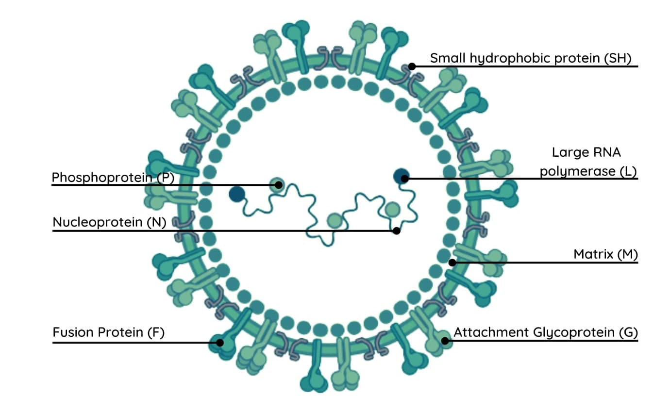
Figure 3: Virion structure of respiratory syncytical virus (RSV)
The RSV virion structure (Figure 3) consists of the nucleoprotein (N), phosphoprotein (P) and large RNA polymerase (L) proteins bound to the RNA strand in the centre of the virion. The two abundant glycoproteins present on the surface of the protective RSV virion envelope are the receptor attachment glycoproteins (G) and fusion (F) proteins. The G and F proteins are critical for initiating the RSV respiratory infection, the G protein is highly glycosylated and targets ciliated epithelial cells, and the F protein enables viral and host cell membrane fusion, leading to the production of new RSV virions. It is the F protein that has been the major target in the development anti-viral drugs to prevent the spread of RSV infections.17,18
Read on to find out more about how we can utilise the differences between each virus to test for COVID-19, flu and RSV.
How do you know if an individual has COVID-19, Flu or RSV?
COVID-19, flu and RSV all share similar symptoms which makes it difficult to know which infection an individual may have at any one time; however, it is important to be able to distinguish between them to avoid inappropriate treatments and reduce the rate of viral transmission. The only way to determine which respiratory virus is causing an infection is to take a highly sensitive real-time genetic test that can accurately differentiate each virus as well as identify cases of co-infection.
Combined COVID-19, flu and RSV testing kits often involve multiplex real-time quantitative reverse transcription PCR (qRT-PCR), which is a highly sensitive and specific assay that uses multiple primers in a single reaction to amplify different nucleic acid sequences simultaneously; by targeting multiple sequences in one single reaction, additional information is gained about the biological sample as well as saving time, money and resources.
References:
- The Royal Society. The SARS-CoV-2 genome: variation, implication, and application. The SARS-CoV-2 Genome. 2020:1-10.
- Bouvier NM & Palese P. The biology of influenza viruses. Vaccine. 2008;26(4):49-53.
- Ali S, Wani JA, Amir S, Tabassum S, Majid S, Eachkoti R, et al. Covid-19: a novel challenge to human immune genetic machinery. Immunogenetics: A Molecular and Clinical Overview. 2022;309-319.
- Wang MY, Zhao R, Gao LJ, Gao XF, Wang DP, Cao JM. SARS-CoV-2: structure, biology, and structure-based therapeutics development. Frontiers in cellular and infection microbiology. 2020;10:587269.
- Wang H, Li X, Li T, Zhang S, Wang L, Wu X, et al. The genetic sequence, origin, and diagnosis of SARS-CoV-2. European Journal of Clinical Microbiology & Infectious Diseases. 2020;39:587269.
- Boopathi S, Poma AB, Kolandaivel P. Novel 2019 coronavirus structure, mechanism of action, antiviral drug promises and rule out against its treatment. Journal of Biomolecular Structure and Dynamics. 2021;39(9):3409-18.
- Zawilska JB, Lagodzinski A, Berezinska M. COVID-19: from the structure and replication cycle of SARS-CoV-2 to its disease symptoms and treatment. Journal of Physiology & Pharmacology. 2021;72(4):479-501.
- Subbarao K, Mahanty S. Respiratory virus infections: understanding COVID-19. Immunity. 2020;52(6):905-9.
- World Health Organization. 2021. Tracking SARS-CoV-2 variants (who.int)
- Jia Z & Gong W. Will mutations in the spike protein of SARS-CoV-2 lead to the failure of COVID-19 vaccines? Journal of Korean medical science. 2021;36(18):124.
- Kesson AM. Respiratory virus infections. Paediatric Respiratory Reviews. 2007;8(3):240-8.
- Saxena SK, Haikerwal A, Gadugu S, Bhatt ML. Complementary and alternative medicine in alliance with conventional medicine for dengue therapeutics and prevention. Future Virology. 2017;12(8):399-402.
- Zhou B, Wentworth DE. Influenza A virus molecular virology techniques. Influenza virus: methods and protocols. 2012;865:175-92.
- Borau MS, Stertz S. Entry of influenza A virus into host cells—Recent progress and remaining challenges. Current Opinion in Virology. 2021;48:23-9.
- Joyce MG, Zhang B, Ou L, Chen M, Chuang GY, Druz A, et al. Iterative structure-based improvement of a fusion-glycoprotein vaccine against RSV. Nature structural & molecular biology. 2016;23(9):811-20.
- Lambert L, Sagfors AM, Openshaw PJ, Culley FJ. Immunity to RSV in early-life. Frontiers in immunology. 2014;5:112039.
- Mlinaric-Galinovic G. Respiratory syncytial virus Part I: From genome to proteome analysis. Virus Research. 2010;153(2):247-57.
- McLellan JS, Ray WC, Peeples ME. Structure and function of respiratory syncytial virus surface glycoproteins. Challenges and Opportunities for Respiratory Syncytial Virus Vaccines. Current Topics in Microbiology and Immunology. 2013;372:83-104.
